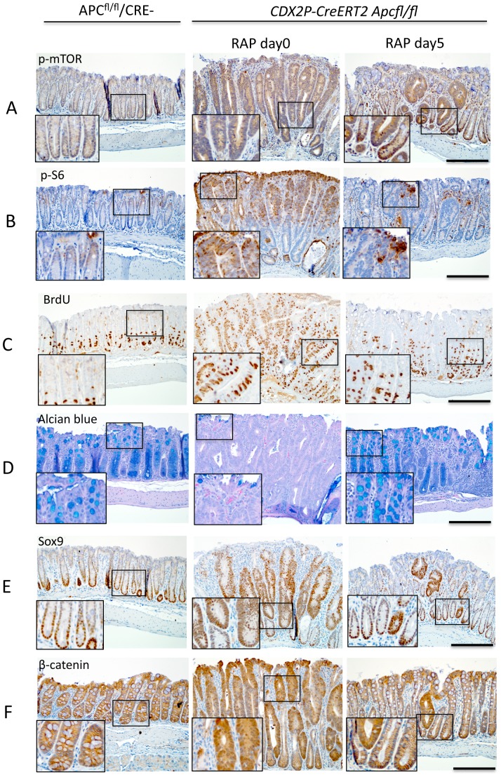
Figure 6. A brief course of RAP treatment decreases markers of proliferation and increases markers of differentiation.
CDX2P-CreERT2 Apcfl/fl mice were injected with TAM and monitored until polyposis was seen endoscopically, then the mice were treated with vehicle or RAP at 3 mg/kg concentration for periods of one, three, or five days (N = 5 per timepoint). (A) and (B) Immunostaining of distal colons from these mice or Cre negative Apcfl/fl mice for phospho-mTOR and phospho-S6. Both phospho-mTOR and phospho-S6 show increased expression in mouse polyps due to Apc inactivation, which is suppressed by 5 days of RAP treatment. (C) Immunostaining of BrdU showed increased incorporation of BrdU in mouse polyps and RAP partially reversed the effect. (D) Alcian blue staining shows loss of most of the differentiated goblet cells in mouse polyps and RAP restore the goblet cell differentiation. (E) Immunostaining shows increased Sox9 (a cell fate marker) expression in polyps and RAP decreased the number of cells expressing Sox9. (F) Immunostaining shows increased nuclear and cytoplastic expression of β-catenin in polyps and RAP treatment for 5 days decreased the expression. Scale bars = 100 µm. The inserts are at two times higher magnification than the original.

