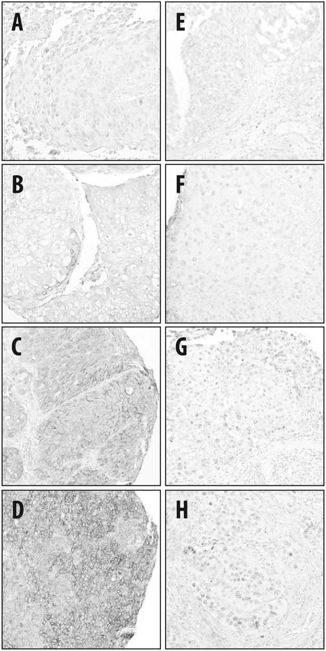
Figure 2.
Representative immunohistochemical staining of tumor samples for (A–D) ribonucleoside reductase subunit M1 and (E–H) excision repair cross-complementation group 1. The staining intensity of the tumor cells was assessed as (A and E) 0, no staining; (B and F) 1, weak; (C and G) 2, moderate; and (D and H) 3, strong staining.

