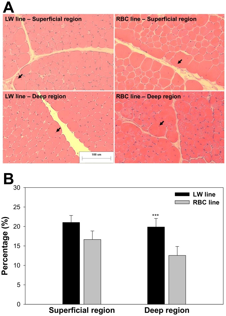
Figure 5. Muscle fiber cross-sections and centered nuclei in the pectoralis major muscle between the low weight (LW) and random bred control (RBC) quail lines at 42 d post-hatch that were stained for hematoxylin and eosin (A); Comparison of centered nuclei percentage relative to total counted nuclei between LW and RBC quail lines at 42 d post-hatch (B).
Arrows indicate centered nuclei. Bars indicate standard errors. Level of significance: *** P<0.001.

