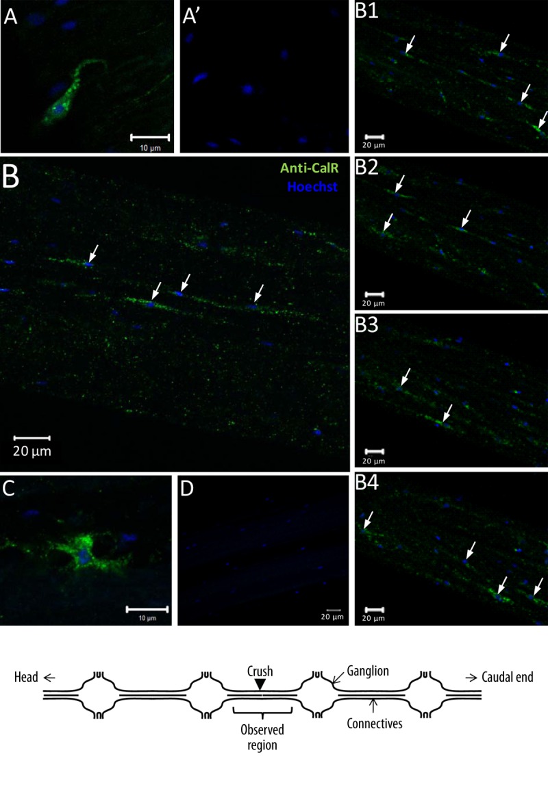
Figure 3.

Hmcalr mRNA and HmCalR protein distribution in isolated segments of leech nervous system (see the diagram below). (A) Fluorescence in situ hybridization on leech connective. Confocal microscopy images showed hmcalr mRNA localization (green) in microglial cells using antisense riboprobes. (A’) Sense probes were used as negative controls. (B and C) Fluorescent immunostaining of HmCalR protein in leech CNS detected at T=0 (B) or T24h post-lesion (C) using rabbit polyclonal anti-human calreticulin antibodies showed immunopositive signal (green) for some microglial cells (arrows). (B1–B4) Immunostaining analyses of successive focal plans in the connective tissues at T=0 allowed the observation of more positive microglial cells (arrows). (D) No immunostaining was observed using secondary antibodies alone as negative control. In any experiment, microglial cell nuclei (blue) were counterstained with Hoechst fluorescent dye.
