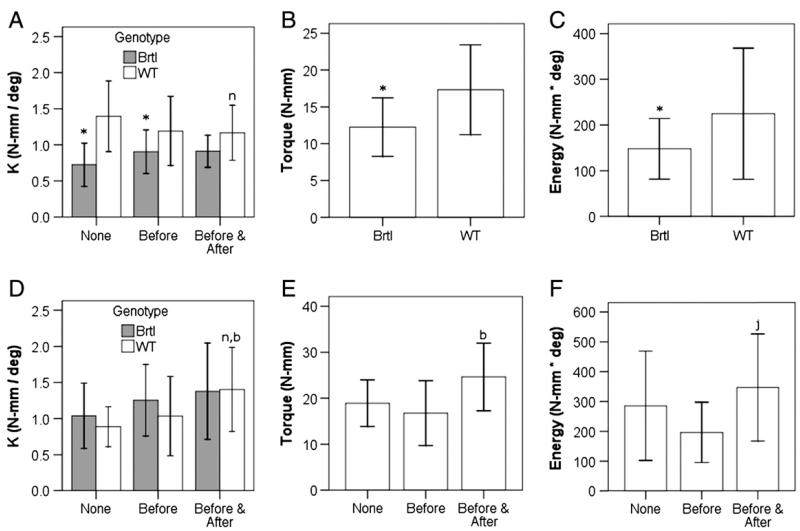
Fig. 4.
Biomechanical changes in fracture calluses based on genotypic and treatment protocol variations at 3 weeks of healing (A–C) and 5 weeks of healing (D–F). Stiffness (A, D), torque at failure (B, E) and energy to failure (C, F) are shown. Notations indicate significance (p < 0.05) with respect to no alendronate treatment (n), alendronate treatment before fracture (b), or between the genotypes (*). In (F), mice that received alendronate before and after fracture compared with those with alendronate before fracture (j, p = 0.053). Genotypes are compared where indicated in A, B, C, and D and are pooled in E and F to compare treatment protocols.

