
Figure 1. Comparison of Cellular Phenotypes within the Venous Neointima in Stenotic AVF (only) and Vein Collected at the Time of Surgical Access Creation.
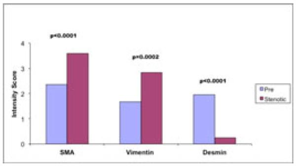
The predominant cellular phenotype within the neointima of vein samples collected at time of new access surgery (pre) were SMA (+), Desmin (+) contractile smooth muscle cells, while in vein segments from stenotic AVF were predominately, SMA(+), vimentin (+) myofibroblasts.