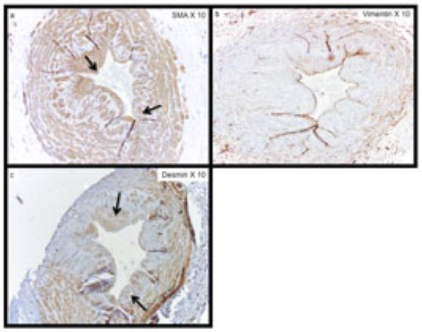
Figure 2. Cellular Phenotyping of Neointimal Cells from Representative Vein Samples Collected at Time of New Surgery.

The expression of (a) α-SMA, (b) vimentin, and (c) desmin within sequential sections of a patient with pre-existing neointimal hyperplasia. The majority of cells within the neointima are SMA (+), vimentin (−), and desmin (+) contractile smooth muscle cells. Arrows show representative areas with SMA(+) and desmin (+) staining within the neointima. Note panel (b) with very little vimentin staining.
