Fig. 6.
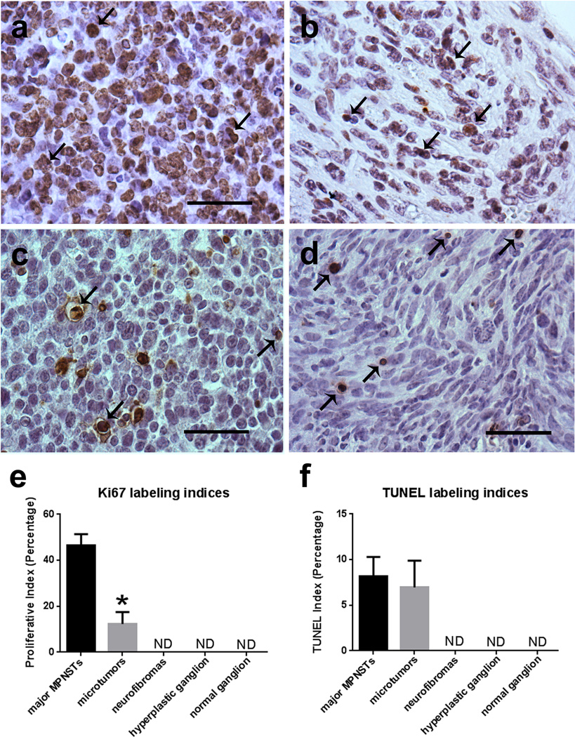
Ki67 labeling indices in the microtumors occurring in P0-GGFβ3; Trp53+/− mice are higher than seen in neurofibromas and non-neoplastic ganglia but lower than is seen in the larger tumors present in these animals. a, b: Major MPNST (a) and micro-MPNST (b) stained for the proliferative marker Ki67 shown side-by-side for comparison. c, d: DNA fragmentation in major MPNSTs (c) and microtumors (d), as labeled via TUNEL. e: Quantification of Ki67 labeling in major MPNSTs, micro-MPNSTs (microtumors), neurofibromas, neoplastic ganglia, and nonneoplastic ganglia demonstrates a significant difference between major MPNSTs and micro-MPNSTs (p<0.0001; n=3 animals per specimen type). f: Quantification of TUNEL labeling in major MPNSTs and micro-MPNSTs, neurofibromas, neoplastic ganglia, and non-neoplastic ganglia. Scale bars in A–D, 50µm.

