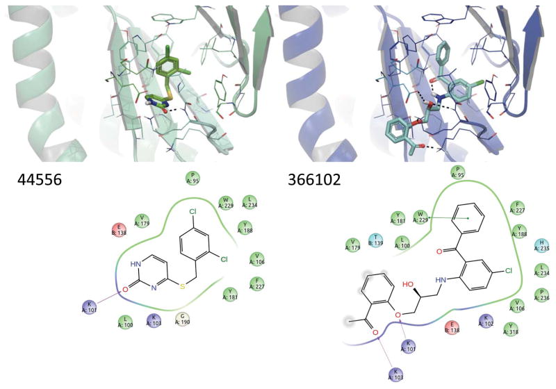
Figure 10.
Proposed binding modes of the 2 confirmed HIV-1 RT inhibitors that were active in enzyme and cell-based assays. Active compounds are shown in predicted poses based on docking into 1RT4 (top). Protein backbone is depicted as ribbons, and residues within 5 Å of the binding site are depicted as sticks. Intermolecular and intramolecular hydrogen bonding are denoted with black dashed lines. Two-dimensional ligand interaction diagrams (bottom) indicate predicted proximal residues for each of the inhibitors.

