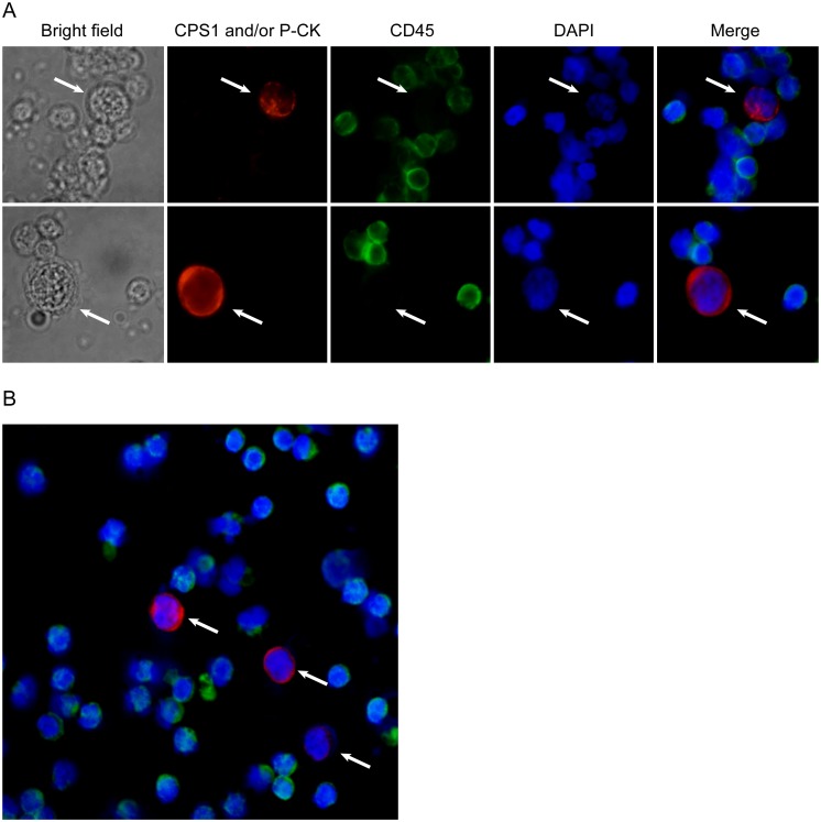
Figure 3. CTCs (indicated by arrow) detected in blood from patients with HCC by the current 3-antibody-based method (magnification, ×200).
(A) A large cell with a morphologically intact DAPI-stained nucleus (blue), CPS1 or/and P-CK (red) positive and CD45 (green) negative was considered a HCC CTC. (B) Several CTCs observed in a same field of view.

