
Figure 1. Sp4 is phosphorylated in response to resting membrane potential.
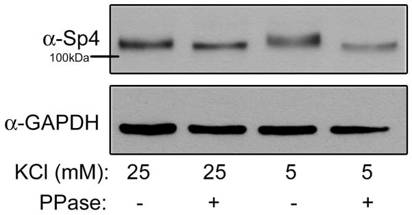
CG neurons were exposed to 25mM (depolarizing) or 5mM (resting) KCl for one hour. Protein extracts were treated with or without a protein phosphatase (PPase), separated by 6% PAGE or 10% PAGE and analyzed by Western blot using antibodies recognizing Sp4 and GAPDH as a loading control. Shown is a representative immunoblot of an experiment performed 5 times.