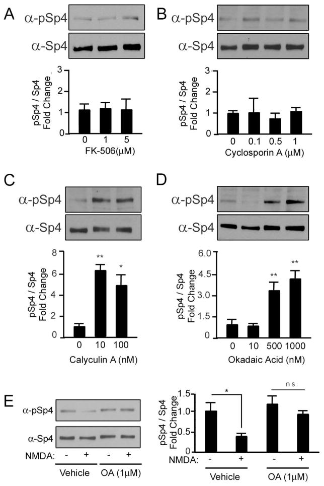
Figure 5. The PP1/PP2A phosphatase reduces Sp4 phosphorylation at S770.
CG neurons were treated with the calcineurin phosphatase inhibitors FK-506 (A) or Cyclosporin A (B) or the PP1/PP2A phosphatase inhibitors Calyculin A (C) or Okadaic Acid (D) at the indicated concentrations for one hour and the levels of phospho-Sp4 S770 relative to total Sp4 were analyzed by Western blot and quantified. N=3–5, *p<0.05, **p<0.01; Calyculin A: ANOVA (F=16.20; p=0.0038); Okadaic acid: ANOVA (F= 6.225; p=0.0018). E. CG neurons were stimulated with 100μM NMDA in the presence of vehicle or 1μM Okadaic acid for one hour, and Sp4 phosphorylation at S770 relative to total Sp4 was determined by Western blot. Left - representative immunoblot. Right - quantification. N=3, *p<0.05, n.s., not significant, unpaired T-test (p=0.0183).

