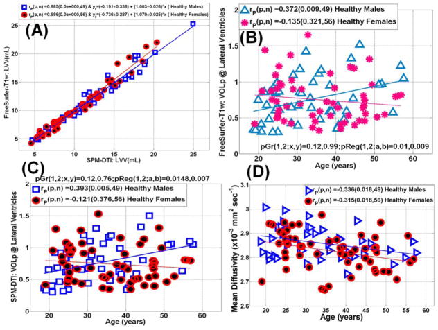
Figure 4.
(A) Scatter plot and linear regression of the lateral ventricular volume obtained using the DTI segmentation and FreeSurfer (see Table 1). (B) A scatter plot and regression analysis of the sensitivity of the normalized LV-to-intracranial volume percentage obtained using FreeSurfer to age in both men and women, note the significant increase in LV volume in men. (C) A scatter plot and regression analysis of the sensitivity of the normalized LV-to-intracranial volume percentage obtain using the DTI-based method to age in both men and women (compare the trends in men and women to those obtained using FreeSurfer Fig. 2C) (D) Scattter and regression analysis of lateral ventricular mean diffusivity in men and women.

