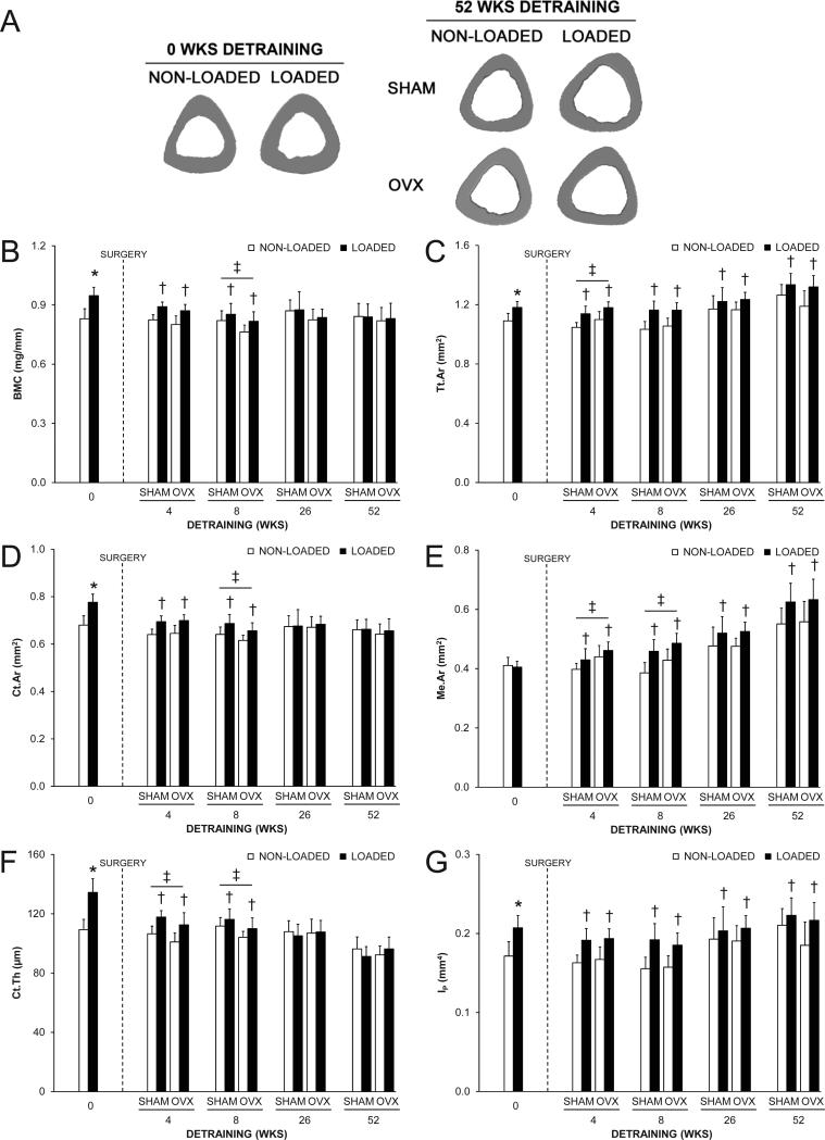
Fig. 2.
The effect of loading and surgery at select detraining time points on the midshaft tibia. A) Representative micro-CT tomographic images of the midshaft tibia in non-loaded and loaded bones from the 0 and 52 wks detraining groups. Loading increased total (Tt.Ar) and cortical (Ct.Ar) areas, and cortical thickness (Ct.Th), as evident in the 0 wks detraining group. The loading-induced increase in Tt.Ar persisted in the 52 wks detraining group in both SHAM and OVX animals. B) Bone mineral content (BMC); C) Tt.Ar; D) Ct.Ar; E) medullary area (Me.Ar); F) Ct.Th and G) polar moment of inertia (IP) at the midshaft tibia as select detraining time points. Loading increased BMC, Tt.Ar, Ct.Ar, Ct.Th and IP, as assessed in the 0 wks detraining group (*p < 0.05). There were no statistical interactions between loading and surgery in any detraining time point group. Loaded tibias had more BMC, Ct.Ar and Ct.Th in the 4 and 8 wks detraining groups and more Tt.Ar, Me.Ar and IP in each detraining time point group than non-loaded tibias (†p < 0.05 for loading main effect). OVX animals had more Tt.Ar and Me.Ar, and less Ct.Th than SHAM animals in the 4 wks detraining group, and less BMC, Ct.Ar and Ct.Th, and more Me.Ar than SHAM animals in the 8 wks detraining group, (‡p < 0.05 for surgery main effect). Data represent body mass corrected means ± SD.

