
Figure 2.
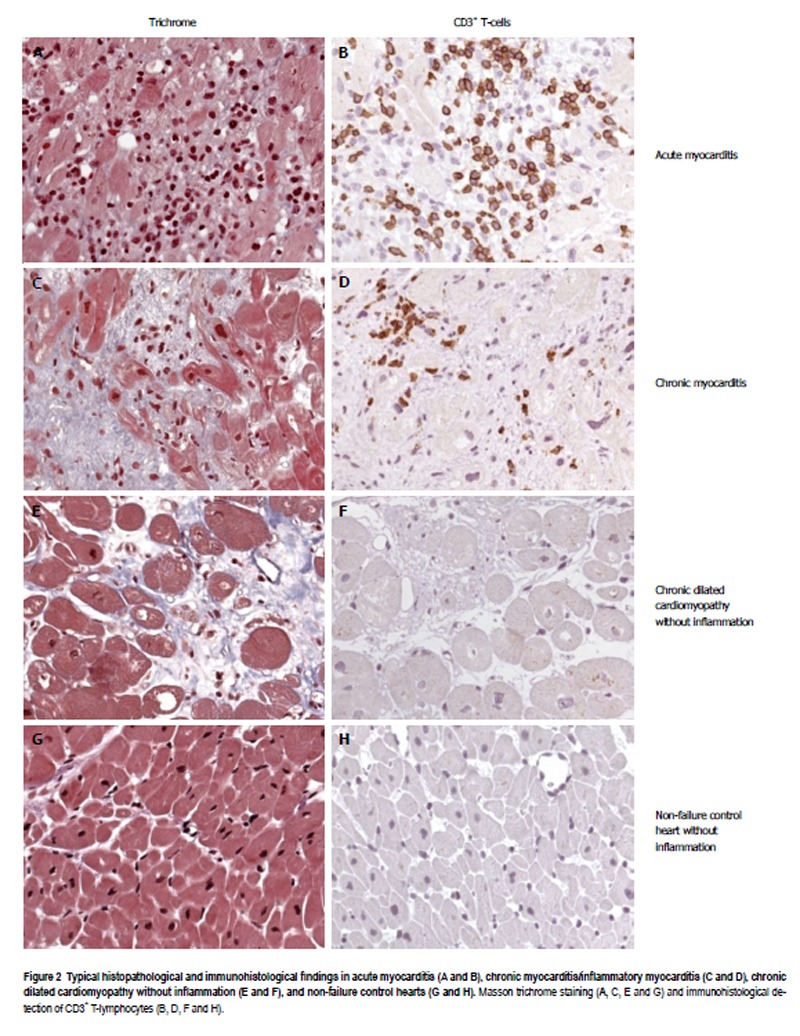
Typical histopathological and immunohistological findings in acute myocarditis (A and B), chronic myocarditis/inflammatory myocarditis (C and D), chronic dilated cardiomyopathy without inflammation (E and F), and non-failure control hearts (G and H). Masson trichrome staining (A, C, E and G) and immunohistological detection of CD3+ T-lymphocytes (B, D, F and H).