
Fig. 1.
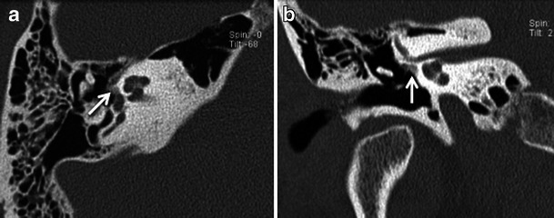
Axial (a) and coronal (b) HRCT images of the right temporal bone in an adult patient with right-sided CHL. A hypodense demineralised plaque (arrow) is noted in the region of the fissula ante fenestram in keeping with fenestral otosclerosis
Official websites use .gov
A
.gov website belongs to an official
government organization in the United States.
Secure .gov websites use HTTPS
A lock (
) or https:// means you've safely
connected to the .gov website. Share sensitive
information only on official, secure websites.
Axial (a) and coronal (b) HRCT images of the right temporal bone in an adult patient with right-sided CHL. A hypodense demineralised plaque (arrow) is noted in the region of the fissula ante fenestram in keeping with fenestral otosclerosis