Fig. 10.

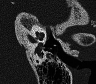
Para-axial HRCT image of the left temporal bone in a patient with severe vertigo, post stapedectomy. The stapes prosthesis is dislocated and lies partly within the vestibule (arrow)

Para-axial HRCT image of the left temporal bone in a patient with severe vertigo, post stapedectomy. The stapes prosthesis is dislocated and lies partly within the vestibule (arrow)