Fig. 13.

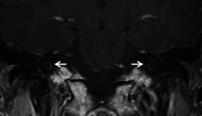
Coronal contrast-enhanced MR image in a patient with left-sided SNHL. Bilateral pericochlear ring-like enhancement (arrow) is suggestive of bilateral cochlear otosclerosis, which was further proven by HRCT

Coronal contrast-enhanced MR image in a patient with left-sided SNHL. Bilateral pericochlear ring-like enhancement (arrow) is suggestive of bilateral cochlear otosclerosis, which was further proven by HRCT