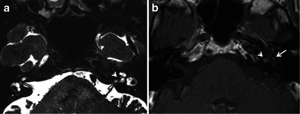
Fig. 14.

a Axial CISS MR image of the skull base in an adult patient with left-sided SNHL. A small hypointense filling defect is seen in the left internal auditory canal (arrowhead), which may be suggestive of an acoustic neuroma. b Axial contrast-enhanced MR image of the same patient as (a), at the same level. The previously noted filling defect in the left internal auditory canal shows post-contrast enhancement, which indicates a small acoustic neuroma (arrowhead). There is also a suggestion of enhancement in the left pericochlear region and in the region of the left fissula ante fenestram (arrow), which suggests associated otosclerosis is present
