Fig. 4.

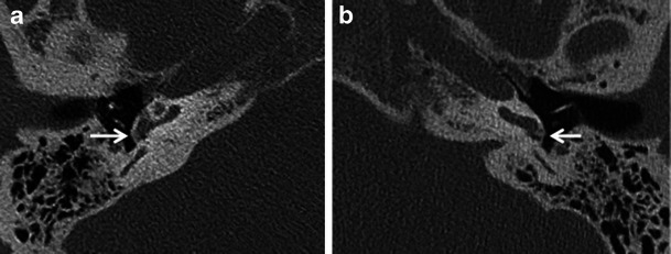
Axial HRCT images of the right (a) and left (b) temporal bone in a patient with bilateral fenestral otosclerosis. Otosclerotic plaques are noted causing bilateral round window narrowing (arrows), right more than left

Axial HRCT images of the right (a) and left (b) temporal bone in a patient with bilateral fenestral otosclerosis. Otosclerotic plaques are noted causing bilateral round window narrowing (arrows), right more than left