
Fig. 7.
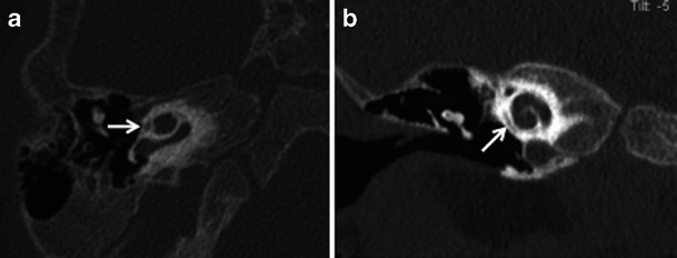
Axial (a) and coronal (b) HRCT images of the right temporal bone in a child with suspected left SNHL. The small lucency seen around the cochlea (arrow) on both the axial and coronal images is in keeping with a cochlear cleft (normal variant)
Official websites use .gov
A
.gov website belongs to an official
government organization in the United States.
Secure .gov websites use HTTPS
A lock (
) or https:// means you've safely
connected to the .gov website. Share sensitive
information only on official, secure websites.
Axial (a) and coronal (b) HRCT images of the right temporal bone in a child with suspected left SNHL. The small lucency seen around the cochlea (arrow) on both the axial and coronal images is in keeping with a cochlear cleft (normal variant)