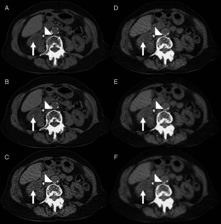
Fig. 4.

A 48-year-old female patient (BMI = 25.6) with acute right ureteric colic. a and b Axial conventional dose (5.1 mSv) non-contrast CT image reconstructed with 40 % ASiR (a) and 100 % FBP (b) showing an 8-mm right ureteric calculus (arrowhead) and an additional 2-mm renal calculus in the right lower pole (arrow) that was not prospectively detected on the low-dose images. c-f Axial low-dose (0.56 mSv) non-contrast CT image reconstructed with 100 % FBP (c), 40 % ASiR (d), 70 % ASiR (e) and 90 % ASiR (f) showing the obstructing 8-mm calculus in the proximal right ureter (arrowhead). The location of the smaller 2.1-mm calculus is also demonstrated (arrow). Note the ‘over smoothening’ of the soft tissues on the 90 % ASiR image compared to the other low-dose reconstructions
