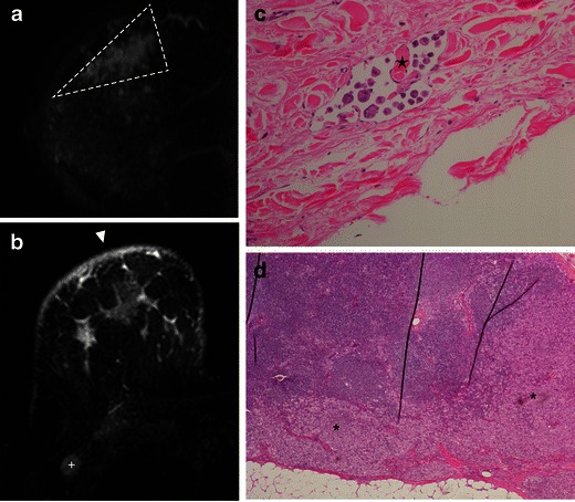
Fig. 10.

A 56-year-old woman. a MR T1-post-contrast subtracted sagittal image showing a non-mass enhancement, with segmental distribution, in an extension larger than seen on mammogram view (dashed line). b) MR—axial STIR demonstrates cutaneous oedema (arrowhead) and axilar adenopathy (+). c H-E ×200, showing tumour embolisation in a dermal lymphatic (star). d Sentinel lymph node biopsy with cortical invasion (*), in H-E stain, ×40
