
Fig. 8.
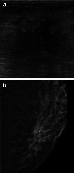
A 73-year-old woman with ILC. a US image depicts an irregular, hypoechoic mass, with indistinct margins and distal attenuation of sound. b Magnified cranio-caudal view also shows an irregular mass associated to microcalcifications
Official websites use .gov
A
.gov website belongs to an official
government organization in the United States.
Secure .gov websites use HTTPS
A lock (
) or https:// means you've safely
connected to the .gov website. Share sensitive
information only on official, secure websites.
A 73-year-old woman with ILC. a US image depicts an irregular, hypoechoic mass, with indistinct margins and distal attenuation of sound. b Magnified cranio-caudal view also shows an irregular mass associated to microcalcifications