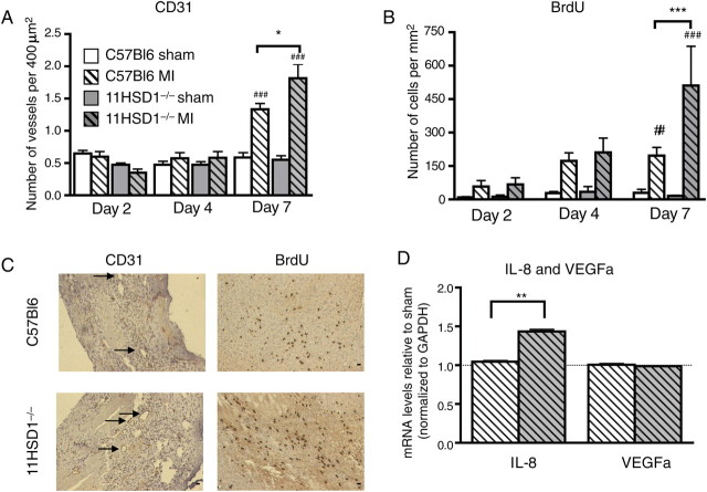
Figure 3.
Vessel density and cell proliferation during infarct healing. (A) CD31-positive vessels <200 μm in diameter in the LV, expressed per 400 μm2. (B) Nuclei positive for BrdU incorporation in the LV, expressed per mm2. (C) Representative sections of infarct border at 7 days after infarction, arrows point to CD31-positive vessels. (D) Interleukin 8 (IL-8) and vascular endothelial growth factor α (VEGFα) mRNA expression levels in heart tissue normalized to GAPDH housekeeping gene at 7 days after MI. n = 8, C57BL/6 sham; n = 12, C57BL/6 MI; n = 4, 11βHSD1−/− sham; n = 6, 11βHSD1−/− MI; for RT–PCR n = 6 per group. #P < 0.05, ###P < 0.001 (sham versus MI). *P < 0.05, **P < 0.01 (C57BL/6 versus 11βHSD1−/−). Scale bar, 10 μm.

