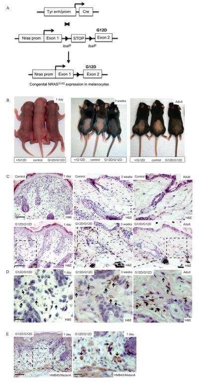
Figure 1. NRASG12D induces skin pigmentation and congenital nevi.
(A) Schematic representation of the conditional-inducible approach used to express NRASG12D in embryonic mouse melanocytes. A tyrosinase gene enhancer/promoter construct (Tyr enh/prom) was used to express Cre-recombinase (Cre) in melanocytes from embryonic day ~10.5 (38). NRASG12D was expressed from the endogenous mouse Nras gene using a conditional-inducible targeted allele in which exon 2 is mutated to introduce the G12D mutation (32). The loxP-STOP-loxP cassette blocks NRASG12D expression, but its removal by Cre-recombinase releases the block on expression.
(B) Photographs showing skin pigmentation in control, Nras+/LSL-G12D;Tyr::CreA/° (+/G12D), and NrasLSL-G12D/LSL-G12D;Tyr::CreA/° (G12D/G12D) mice, at 1 day, 3 weeks, and in adulthood.
(C) Upper panels: photomicrographs of H&E stained skin in 1 day old, 3 week old and adult mouse skin. Scalebar = 200μm. Lower panels: low power photomicrographs of H&E stained skin in 1 day old, 3 week old and adult NrasLSL-G12D/LSL-G12D;Tyr::CreA/° (G12D/G12D) mice. Hyperpigmented dendritic melanocytes are visible at low magnification in the 3 week old and adult mice. Scalebar = 200μm. n=6 mice / experimental group.
(D) High power photomicrographs of H&E stained skin from boxed areas in the lower panel from (C) in 1 day old, 3 week old and adult NrasLSL-G12D/LSL-G12D;Tyr::CreA/° (G12D/G12D) mice. Hyperpigmented dendritic melanocytes in the papillary and reticular dermis, and along the hair follicles and adnexal glands are indicated (black arrows) and are sparse in the skin of day 1 old mice, but are prominent in 3 week old and adult mice. Scalebar = 20μm.
(E) Photomicrographs of HMB45/MelanA stained skin from a 1 day old NrasLSL-G12D/LSL- G12D;Tyr::CreA/° (G12D/G12D) mouse, demonstrating the presence of melanocytes (black arrows). The area boxed in the left hand panel is enlarged in the right hand panel. Scale bars = 200μm (left panel) and 20μm (right panel).

