
Fig. 4.
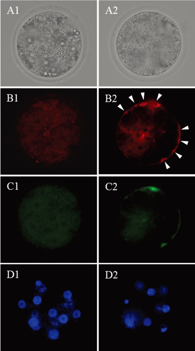
Bright field (A) and fluorescent micrographs (B, C and D) of embryos (72 hpi) derived from the oocytes subjected to 22 (1) and 34 h (2) of IVM culture. B, high-polarized mitochondria stained by JC-1; C, low-polarized mitochondria stained by JC-1; D, nuclei stained by Hoechst 33342. Twelve and 8 nuclei are observed in D1 and 2, respectively. The embryo derived from the oocyte subjected to 22 h of IVM shows high-polarized mitochondria at the periphery of blastomeres (white arrows: B2).