Fig. 2.
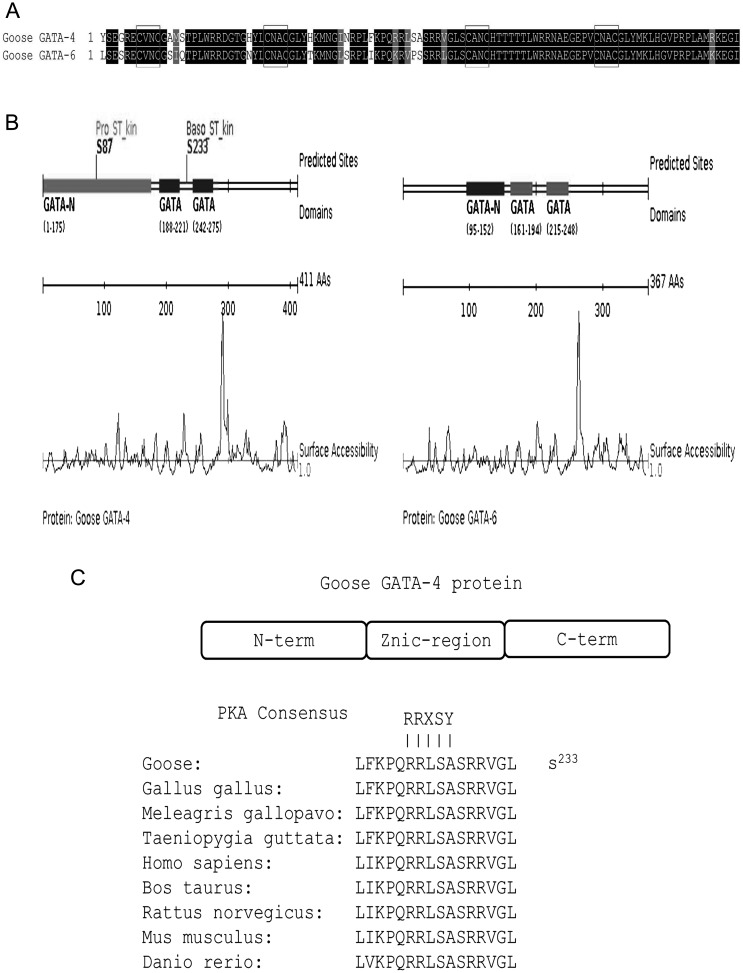
A: Amino acid sequence alignment of the zinc finger domains between goose GATA-4 and GATA-6. The black blocks represent identical residues between the zinc finger domains shown, while the red boxes represent the distinct form (CVNC-X17-CNAC)-X29-(CANC-X17-CNAC). B: The phosphorylation sites in goose GATA-4 and GATA-6 proteins were predicted by Scansite 3. The GATA-4 protein contains two phosphorylation sites located at amino acid positions 87 (S87) and 233 (S233). No sites were found in goose GATA-6. C: Mapping of the phospho-residue reveals that the GATA-4 protein contains a species-conserved PKA consensus phosphorylation site located within the zinc finger region.

