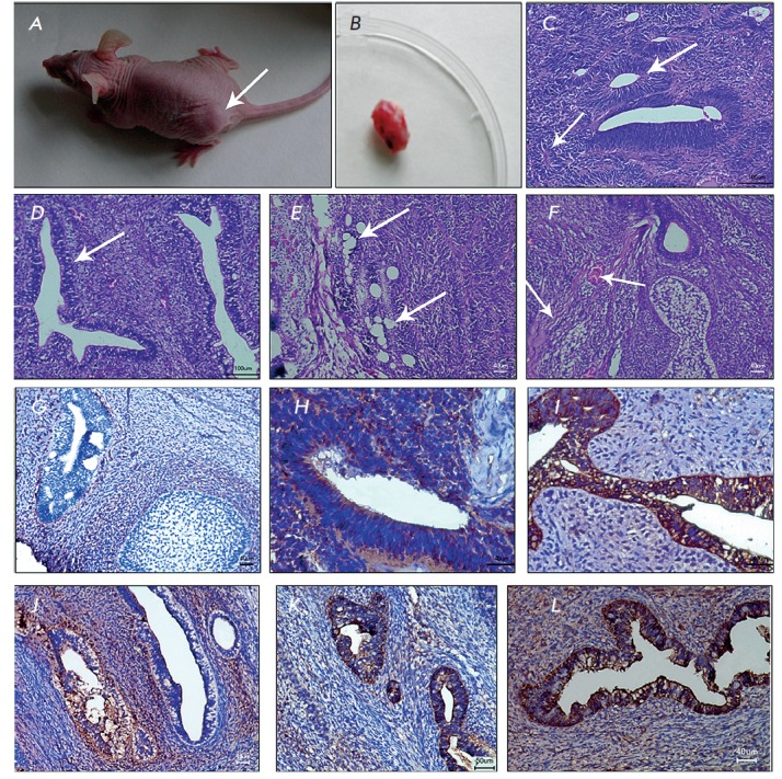
Fig. 4.

Formation of teratoma from iDP cells. The recipient mouse (A) with teratoma (arrow) and the resected teratoma (B) six weeks after subcutaneous injection of iDP cells. Histological sections of hematoxylin and eosinstained teratomas (C–F): neuroglia (neuroepithelial tubules and rosettes, arrows) (C); glandular epithelium (D), mesenchyme (adipose cells, arrows in D; fibers of loose connective tissue and vessels, arrows in F) (E, F); stained with antibodies against: vimentin (G), nestin (H) , pan CK (I) CK8 (J), CK18 (K) and AFP (L)
