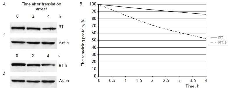
Fig. 3.

Comparison of RT and RT-Ii degradation in the expressing cells. A – Immunoblotting of HeLa cells transfected with pKCMV2RT (1) and pKCMV2RT-Ii (2) sampled at the given time-points after the addition of cycloheximide (100 μg/ml). Blots were stained with anti-RT polyclonal antibodies. To normalize the signal to the total protein content of the loaded samples, the membranes were stripped and re-probed with anti-actin antibodies. B – The kinetics of degradation of RT and RT-Ii
