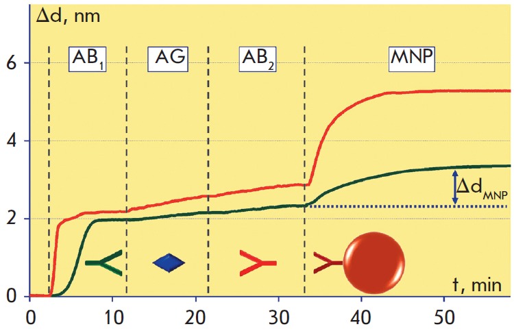
Fig. 8.

Sensograms demonstrating all stages of the magnetic immunoassay on the biotinylated (bottom green curve) and epoxylated (top red curve) surfaces of the sensor chip: AB1 – antibody immobilization; AG – antigen (100 ng/ml) capture by immobilized antibodies; AB2 – recognition by tracer antibodies of another antigen epitope; MNP – association of magnetic nanoparticles with tracer antibodies. PBS washing was performed before each step as indicated with dotted lines
