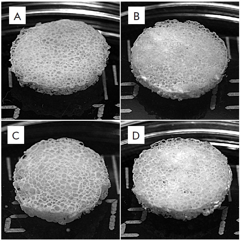
Fig. 1.

Appearance of 3D porous silk fibroin (A) and composite fibroin–gelatin (B), fibroin–hydroxyapatite (C), and fibroin–gelatin–hydroxyapatite (D) scaffolds. Introduction of gelatin and hydroxyapatite into the scaffold structure does not modify its appearance
