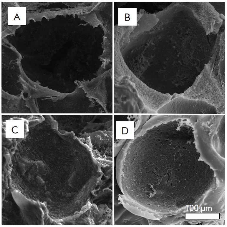
Fig. 2.

Structure of 3D porous silk fibroin (A) and composite fibroin–gelatin (B), fibroin–hydroxyapatite (C), and fibroin–gelatin–hydroxyapatite (D) scaffolds. The images were recorded on a scanning electron microscope. Introduction of gelatin and hydroxyapatite into the scaffold structure does not modify the pore size and the general scaffold structure
