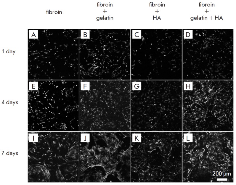
Fig. 4.

GFP-expressing murine embryonic fibroblasts (MEF) on the silk fibroin scaffold (A, E, I), composite fibroin–gelatin scaffold (B, F, J), hydroxyapatite (C, G, K), gelatin and hydroxyapatite (D, H, L) after 1 (A–D), 4 (E–H), and 7 (I–L) days of cultivation. The images show surface projections of the optical sections
