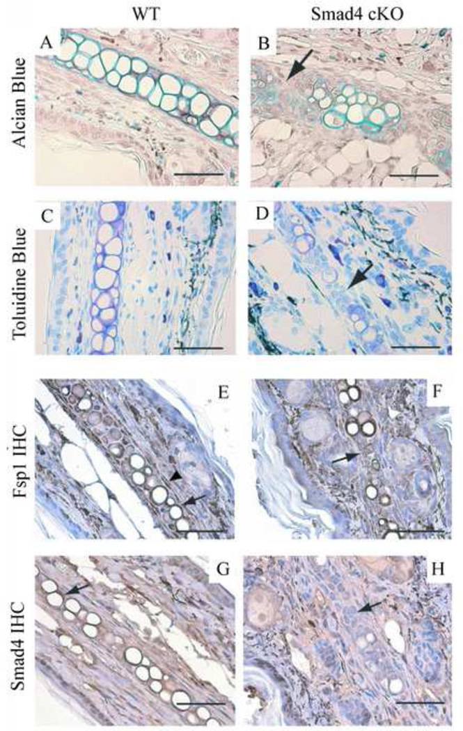
Figure 2. The Smad4 cKO ears are deficient in cartilage.
In the Smad4 cKO ears, there is less cartilage (B: Alcian blue stain: turquoise; D: Toluidine blue stain, purple), especially around the clusters of immature chondrocytes (arrow). WT control stains: A: Alcian blue stain. C: Toluidine blue stain. E-F: Fsp1 labeling. E. In WT ear tissue, Fsp1 is expressed in the fibroblasts (arrowhead) and the chondrocytes (arrow). F. In the Smad4 cKO chondrocytes, Fsp1 is also expressed in the chondrocytes. Arrow points to a cluster of immature chondrocytes that are positive for Fsp1. G-H: Smad4 labeling. G. Smad4 is expressed strongly in the WT ear chondrocytes (arrow). H. The Smad4 cKO ear chondrocytes show insignificant Smad4 staining. Arrow points to a cluster of immature chondrocytes that are negative for Smad4 staining. Scale bar: 50µm.

