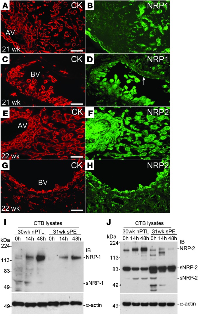
Figure 3. NRP-1 and NRP-2 (protein) expression at the maternal-fetal interface in normal pregnancy and in sPE.
Tissue sections were double stained with anti–cytokeratin-8/18 (CK), which reacts with all trophoblast subpopulations, and anti–NRP-1 or NRP-2. (A and B) NRP-1 expression was detected in association with villous trophoblasts. Within the uterine wall, immunoreactivity associated with invasive CTBs was upregulated as the cells moved from the surface to the deeper regions. (C and D) Endovascular CTBs that lined a maternal blood vessel (BV) were also stained. (E–H) Anti–NRP-2 reacted with trophoblast and nontrophoblast cells in anchoring villi (AV) as well as interstitial and endovascular CTBs. Essentially the same staining patterns, but with weaker intensity, were observed in sPE (data not shown). CTBs were isolated from the placentas of control nPTL cases and from the placentas of women who experienced sPE. (I) Over 48 hours in culture, NRP-1 expression was upregulated in both instances but to a lesser degree in sPE. (J) Control nPTL CTBs also upregulated NRP-2. Expression of this receptor was reduced in sPE and the soluble form was more abundant. (A–J) The data shown are representative of the analysis of a minimum of 3 samples from different placentas. Scale bars: 100 μm. AV, anchoring villi.

