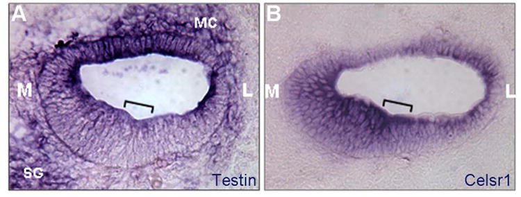
Fig. 3.

Testin is expressed in the inner ear during development.
(A-B) E14.5 cochlear sections probed for Testin (A) or Celsr1 (B). The bracket in each panel marks the developing organ of Corti. M: medial or center of the cochlear spiral; L: Lateral or periphery of the cochlear spiral; MC: mesenchyme cells; SG: spiral ganglion neurons.
