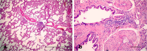
Figure 10.

Photomicrographs of hematotoxylin and eosin-stained sections of the lung in (LTC plus Garlic extract) group, 15 d p.i. (HE stain). (a) Mild interstitial inflammation and mild hyperplasia shown in the bronchiolar lymphoid follicles (Bar = 200 μm); (b) Mild peribronchiolitis manifested by focal areas of round cells accumulating in the wall of bronchioles (Bar = 50 μm).
