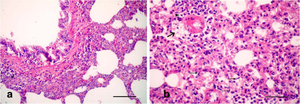
Figure 8.

Photomicrographs of hematotoxylin and eosin-stained sections of the lung in (LTC plus Gensing) group, 21 d p.i. (HE stain). (a) Mild hyperplasia in the small bronchioles with a normal lumen; (b) Alveolitis manifested by edema (arrow) with aggregation of mononuclear cells (arrowhead) around the alveolar blood vessels, with hyperplasia in the alveolar septa, besides pulmonary fibrosis (thin arrows) (All Bars = 50 μm).
