
Figure 1.
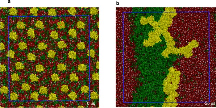
Top view of snapshots from a CGMD simulation of a domain-forming bilayer with asymmetrically bound lipid-modified proteins: (a) the initial setup at 0 μs; (b) the final configurations at 25 μs. DPPC is shown in red, DLiPC in green, cholesterol in white, and protein in yellow. Shown in blue is the actual simulation box, with the region outside being part of the periodic images in each direction. See Figure S1 (SI) for another aggregate from a different simulation.