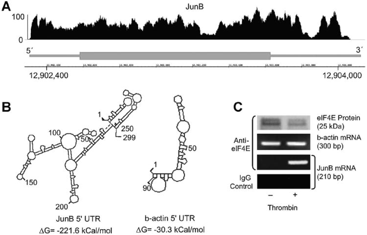
Fig. 6.

Thrombin stimulation induces association of eIF4E and JunB mRNA in human EC. Panel A: Next-generation RNA-sequencing of endothelial cell JunB transcript 5′-UTR. Polyadenylated RNA was isolated from unstimulated EC and was subsequently used for next-generation RNA-sequencing. The JunB gene (bottom portion) is represented by a thick (exon) lines, 5′- and 3′- ends of the gene are labeled accordingly. The position on the chromosome is indicated by the numbers on the x-axis. The diagram above the gene pictorial (top portion) represents EC-derived transcripts that were fragmented, sequenced, and subsequently matched and aligned to JunB using the Integrated Genome Browser (IGB). The y-axis represents the abundance of hits matched to the appropriate part of the gene. Panel B: Predicted folding structures and associated free energy values for the 5′-UTRs of JunB and b-actin (DNASIS, Hitachi Genetic Systems). The predicted secondary structures and free energies of the JunB and β-actin 5′ UTR are illustrated. A free energy value ≤50 kcal/mol suggests that a regulated event to linearize the secondary structure is necessary to efficiently translate the mRNA transcript. Panel C: JunB mRNA is detected only in eIF4E immunoprecipitates derived from thrombin-stimulated EC. Human EC were treated with thrombin (2 U/ml, 2 h). Immunoprecipitates were prepared using anti-eIF4E (or non-immune IgG) antibodies and probed for either β-actin or JunB mRNA by PCR.
