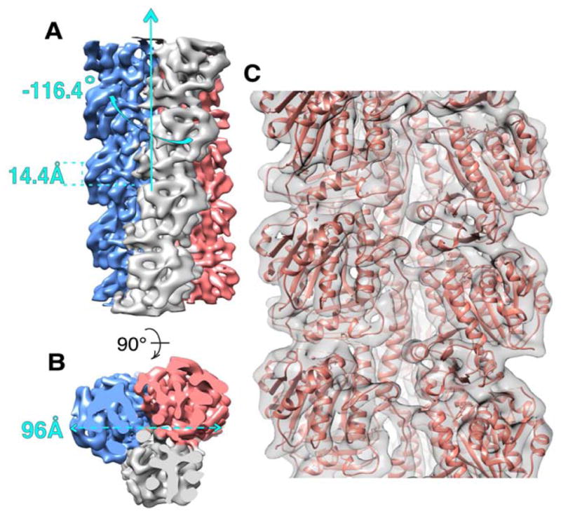
FIGURE 2. Cryo-EM map and pseudo-atomic model of PhuZ201 filament.

(A) and (B) Cryo-EM map of PhuZ201 filament with each protofilament presented in a different color. (A) Map has helical symmetry of −116.4° rotation and 14.4 Å rise per subunit. (B) End-on view of the filament shows that it is a trimer with 96 Å diameter. (C) Pseudo-atomic model of PhuZ201 filament. In gray surface is the cryo-EM density fitted with the atomic models of PhuZ201 in salmon. See also Figure S1 and Movie S1.
