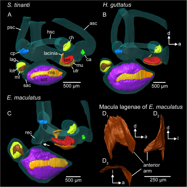
Figure 3.
Spatial orientation and curvature of sensory epithelia (maculae and cristae) in the studied species. While the anterior arm of the macula lagenae is directed upward in S. tinanti(A) and H. guttatus(B), it points anteriorly in E. maculatus(C, D). A 3D model of the macula lagenae of E. maculatus based on histological serial sections illustrates the strong curvature of the anterior arm (D1-D3). In E. maculatus(C), part of the lacinia of the macula utriculi forms a dorsal “roof” above the utricular otolith. The white arrow in (C) marks the thin connection between saccule and the upper part of the inner ear. 3D reconstructions are based on microCT scans (A-C, isotropic voxel size 4 μm) and histological section series (D, voxel size x = y = 0.54 μm; z = 3 μm). a, anterior; ca, crista of the anterior semicircular canal; ch, crista of the horizontal semicircular canal; cp, crista of the posterior semicircular canal; d, dorsal; l, lateral; lag, lagena; lot, lagenar otolith; ml, macula lagenae; ms, macula sacculi; mu, macula utriculi; rec, recessus situated posteriorly to the utricle; sac, saccule; sot, saccular otolith; uot, utricular otolith; utr, utricle. Scale bars, 500 μm (A-C), 250 μm (D).

