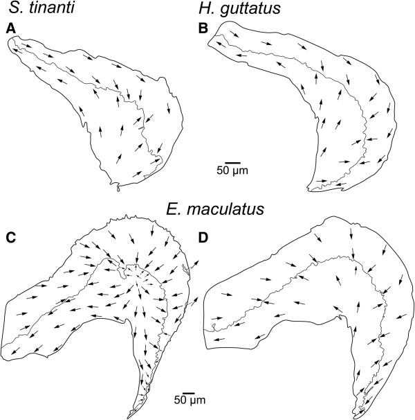
Figure 6.
Interspecific comparison of the orientation patterns of ciliary bundles on the macula lagenae. Ciliary bundles of all species are mainly arranged into two “opposing” groups (A-B, D). In E. maculatus, however, distinct intraspecific variability can be observed as shown in (C)vs.(D). In (C), ciliary bundles in the center of the macula show a radial arrangement. Note that the arrows point into the direction of the kinocilia indicating the orientation of the ciliary bundles in the respective area while the dashed lines separate different orientation groups. Scale bars, 50 μm.

