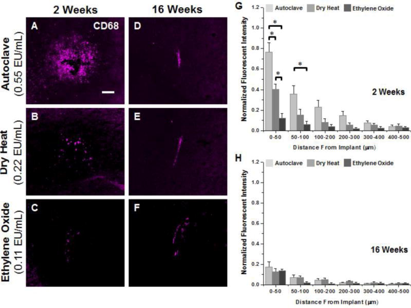
Figure 3.

Representative immunohistochemical images of CD68+ immunoreacivity surrounding microelectrodes sterilized by autoclave, dry heat or ethylene oxide gas at 2 weeks (A–C) and 16 weeks (D–F) post implantation. Quantification of CD68+ immunoreactivity at 2 weeks post implantation (G) shows significantly higher levels between 0–100μm for microelectrodes sterilized by autoclave compared to microelectrodes sterilized by dry heat or ethylene oxide gas. Further, significantly higher CD68+ immunoreactivity was observed for microelectrodes sterilized by dry heat compared to ethylene oxide gas within the first 50μm. At 16 weeks post implantation (H), similar levels of CD68+ immunoreactivity was seen around all implant, regardless of initial endotoxin levels. Scale Bar = 100μm; * p<0.02
