Figure 4.

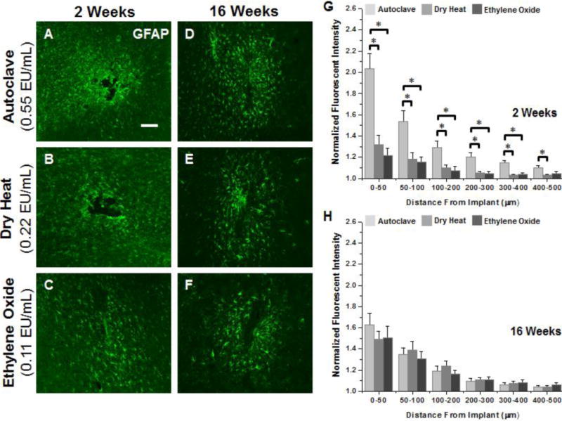
Representative immunohistochemical images of GFAP+ immunoreactivity surrounding microelectrodes sterilized by autoclave, dry heat or ethylene oxide gas at 2 weeks (A–C) and 16 weeks (D–F) post implantation. Quantification of GFAP at 2 weeks (G) shows significantly heightened astrogliosis up to 400μm from the interface for microelectrodes sterilized by autoclave compared to microelectrodes sterilized by dry heat or ethylene oxide gas. At 16 weeks post implantation (H) no difference was seen regardless of initial endotoxin levels. Scale Bar = 100μm; * p<0.02
