Figure 6.

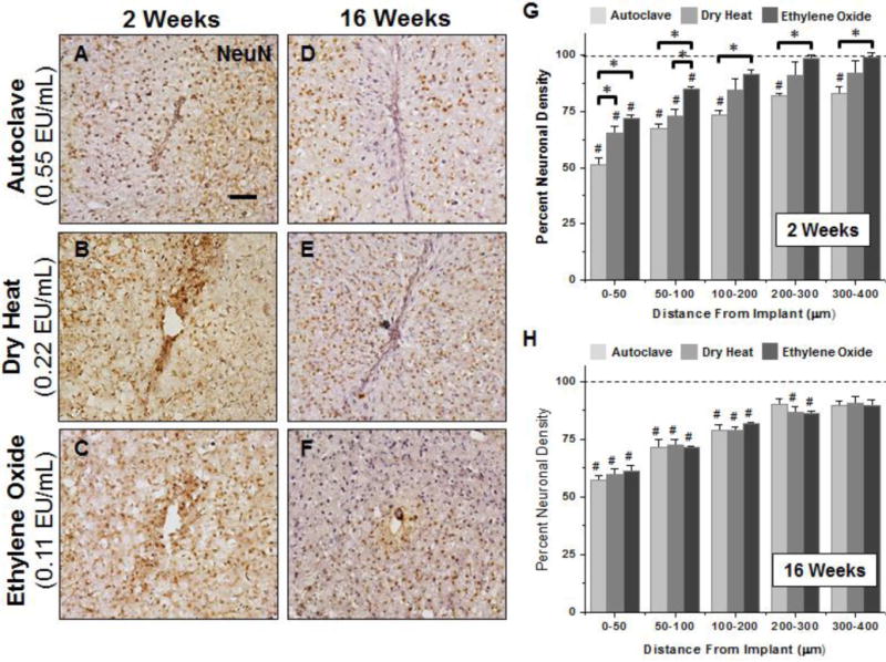
Representative immunohistochemical images of neuronal nuclei density surrounding microelectrodes sterilized by autoclave, dry heat or ethylene oxide gas at 2 weeks (A–C) and 16 weeks (D–F) post implantation. For all binned intervals at 2 weeks (G), microelectrodes sterilized by autoclave had significant reduced neuronal density compared to microelectrodes sterilized by ethylene oxide gas as well as background neuronal density of non-surgical age-matched controls. Further, significant decreases in neuronal densities were seen between microelectrodes sterilized by autoclave compared to microelectrodes sterilized by dry heat and ethylene oxide gas between 0–50μm. At 16 weeks, neuronal density remained significantly lower than background neuronal density up to 200μm, regardless of sterilization method. However, at 16 weeks post implantation we observed no significant difference based on initial endotoxin levels. Scale Bar = 100μm; * p<0.02; # p<0.05
