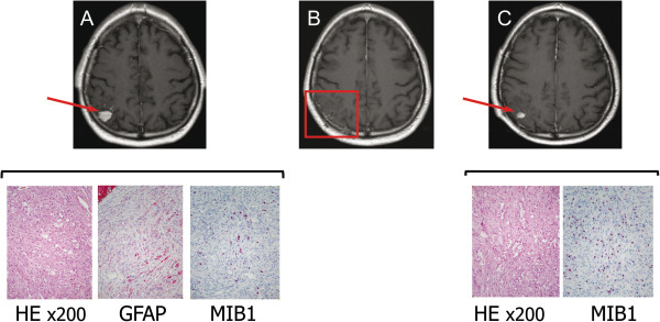
Figure 1.
Immunohistochemical characterization of grade II PXA and associated anaplastic PXA. Upper row: T1-weighted, gadolinium-enhanced axial MRI showing the right parietal PXA at presentation (A), after surgical resection (B) and at the time of local relapse (C). Lower row: Photomicrographs showing histological and immunohistochemical features of the grade II PXA and grade III PXA with anaplastic features (HE, GFAP and MIB-1. The last one showed a higher positivity of 20% in the anaplastic PXA, whereas it was 10% in the grade II PXA).

