
Figure 7. EphA/ephrin signaling is upstream of the generation of the actomyosin cables.
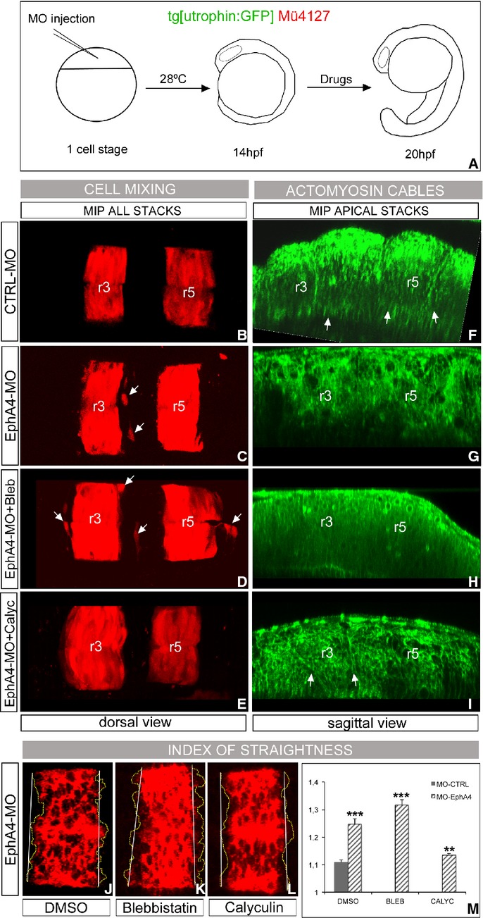
A–I Presence of actomyosin cables and effects in rhombomeric cell segregation. (A) Scheme of the functional experiment: double transgenic Mü4127/Tg[utrophin:GFP] embryos injected with CTRL-MO (B, F) or EphA4a-MO (C–I) at 1- to 2-cell stage, incubated from 14 hpf for 6 h with DMSO (B–G), Blebbistatin (D, H), or Calyculin A (E, I). After the treatment, the degree of cell mixing (B–E) and the presence of actomyosin cables (F–I, see arrows) were assessed. Embryos injected with CTRL-MO behave as control embryos (DMSO) in previous experiments. Note the cell mixing in embryos in which the cable was dismantled (white arrows, in C–D, G–H), and the partial rescue of the cable in EphA4a-MO embryos treated with Calyculin A (white arrows in I) resulting in no cell mixing (E). Dorsal views (B–E) and sagittal-optical views of apical stacks (F–I); in all cases anterior is to the left.
J–M Analysis of the index of straightness (IS) in wt embryos injected with EphA4a-MO at 1- to 2-cell stage, incubated from 14 hpf for 6 h with different pharmacological agents, and assayed for krx20 in situ hybridization. Note the jagged krx20 expression domains upon Blebbistatin treatment, and how the effect of EphA4a-MO is enhanced. IS is partially rescued in morphants upon Calyculin A treatment. Dorsal views with anterior to the left. (M) Quantification of the IS for embryos in experiment (J–L) (dashed bars), and comparison with control embryos (solid bar). ***P < 0.001, **P < 0.005.