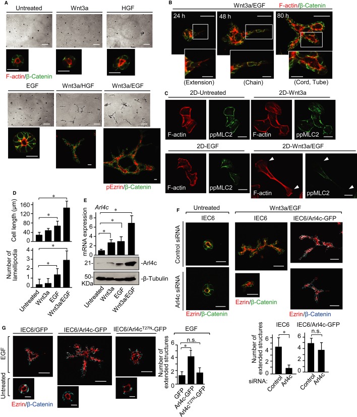
Figure 1. Arl4c expression is required for tube formation of IEC6 cells in 3D culture.
A IEC6 cells were cultured under the indicated conditions for 72 h in 3D Matrigel. Cells were photographed using phase contrast microscopy and stained with the indicated antibodies or phalloidin.
B IEC6 cells were cultured with Wnt3a and epidermal growth factor (Wnt3a/EGF) for 24, 48, or 80 h in 3D Matrigel and stained with anti-β-catenin antibody and phalloidin.
C, D IEC6 cells grown on a Matrigel-coated coverslip (2D culture) were cultured under the indicated conditions for 18 h and stained with the indicated antibody and phalloidin. White arrowheads indicate lamellipodia (C). The length of the long axis and the number of lamellipodia of cells were measured per treatment (n = 50) (D).
E IEC6 cells were stimulated as indicated for 18 h. Real-time PCR analyses for Arl4c mRNA expression were performed. The results are expressed as fold increase compared with Arl4c mRNA levels in untreated cells. Whole lysates were probed with the indicated antibodies.
F IEC6 cells or IEC6 cells stably expressing Arl4c-GFP (IEC6/Arl4c-GFP) were transfected with control or Arl4c siRNA and cultured with or without Wnt3a/EGF for 60 h. The cells were stained with the indicated antibodies. The number of extended structures from multicellular trunks was counted (n = 30).
G IEC6 cells stably expressing GFP, Arl4c-GFP, or Arl4cT27N-GFP were cultured with or without EGF for 60 h and stained with the indicated antibodies.
Data information: Results are shown as the mean ± SE from three independent experiments. Scale bars in (A), 200 μm (top panels) and 20 μm (bottom panels); in (B), 50 μm (top panels) and 20 μm (bottom panels); in (C), 20 μm; in (F), 20 μm (left two panels) and 50 μm (right four panels); in (G), 50 μm (top panels) and 10 μm (bottom panels). *P < 0.01; n.s., not significant.
Source data are available online for this figure.

